
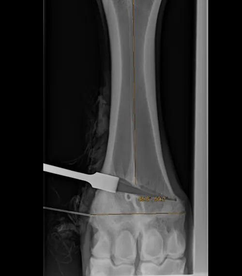
Figure 1: Correction of the left distal metacarpal valgus with osteotomy, plate fixation and Xcyte® implantation.
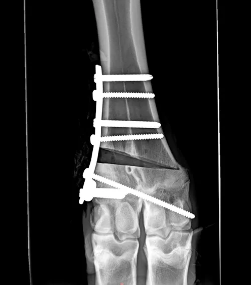
Figure 2: Immediate postop, alignment following osteotomy, fixation and Xcyte implantation.
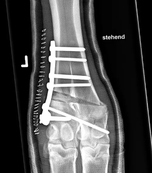
Figure 3: 3 week follow-up showing significant bone growth at the osteotomy site, and stable plate fixation.
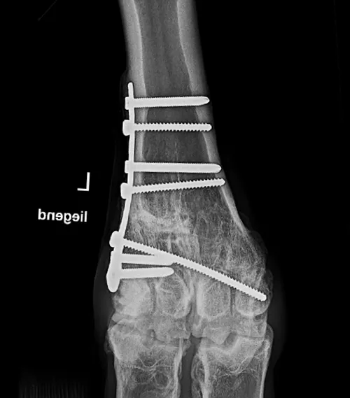
Figure 4: Long-term follow-up confirming osteotomy healing, with maintained alignment and good limb function.
Correction of a congenital valgus malformation of the left thoracic limb
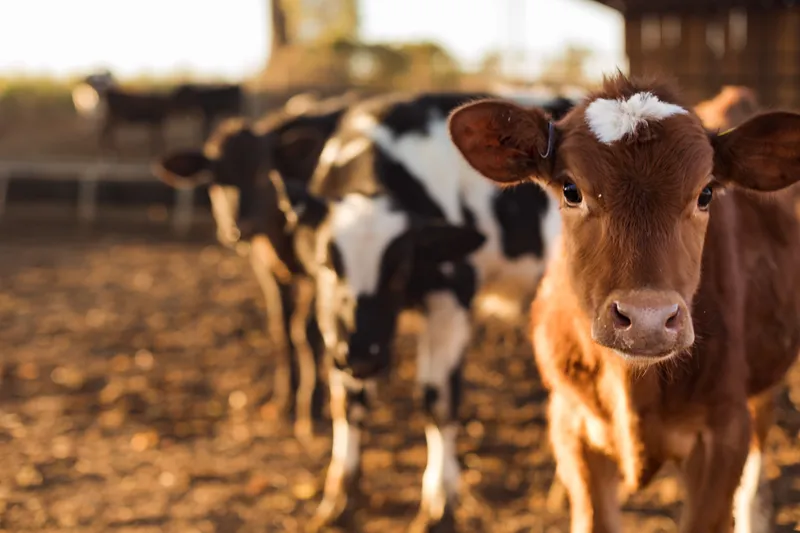
A 9-month-old female Red Holstein calf was presented to Vetsuisse Bern, Switzerland, with a congenital valgus deformity affecting both front limbs, more pronounced on the left side. Clinical examination revealed visible angular deviation and lameness.
Radiographic examination confirmed valgus deviation at the level of the distal metacarpal growth plate, with associated bone and physeal remodeling, as well as bilateral regional soft tissue swelling.
Due to the deformity and associated functional impairment, corrective osteotomy was performed to restore alignment and support long-term mobility. A transposition osteotomy of the left distal metacarpus was performed under radiographic control (Figure 1). The osteotomy was stabilized using a plate and screws, and Xcyte, an osteoinductive bone graft substitute, was inserted into the osteotomy site (Figure 2). A walking cast was applied during surgery to provide additional support during the initial healing period.
Following surgery, the calf recovered well and was able to bear weight with support from the cast. The patient was discharged approximately one month after surgery. Radiographic examination at 3 weeks demonstrated significant bone growth at the osteotomy site, with stable plate fixation (Figure 3).
At long-term follow-up, 33 weeks after surgery, radiographs demonstrated progressive healing and bone remodeling, with only mild residual valgus deviation. Clinically, the calf showed improved limb function, with no visible lameness in the left front limb.